Figure 11.
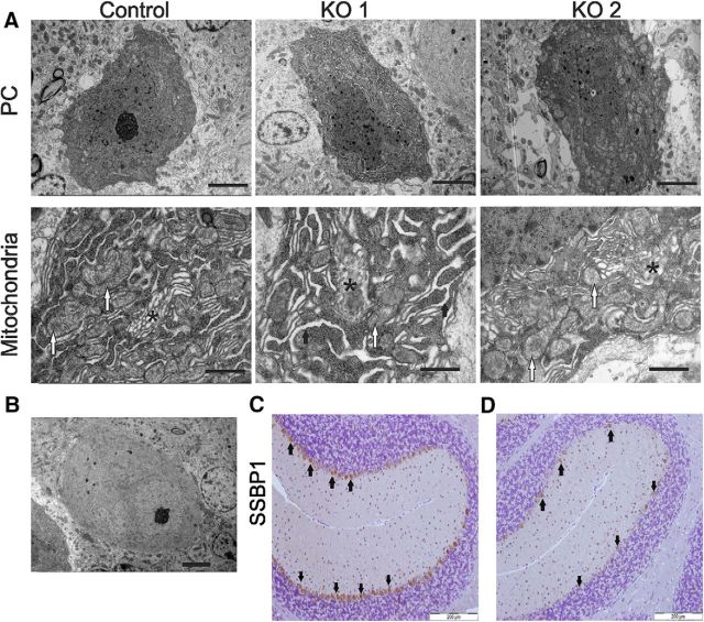
Ultrastructural analysis of PCs in control and Mecr-KO samples at 6 months of age by electron microscopy and assessment of PC mtDNA content. A, Example PC in one KO mouse displays mitochondria with a pale matrix and dilatation of the endoplasmic reticulum (KO 1), whereas the PC of another KO mouse has only mitochondria with pale matrix (KO 2). Scale bars in top row of A, 5 μm. Most of the mitochondria in the KO mouse PCs lack clear cristae and contain a pale matrix (bottom row of A, white arrows). The vesicular Golgi apparatus (*) in KO mice is disorganized. The endoplasmic reticulum is dilated in some of the KO mice at 6 months of age as exemplified here by the KO 1 (thick arrows). Scale bar, lower row A, 1 μm. B, Representative image of a pale PC from a KO mouse. Scale bar, 5 μm. C, D, Comparison of IHC detection of mitochondrial nucleoids identified by anti-single-stranded binding protein (SSBP1) antibody in control (C) and KO mouse (D) cerebella revealed weaker (brown) staining in PCs of the latter, indicating possible reduction of mitochondrial nucleoids in PCs of KO mice at 6 months of age. Scale bar, 200 μm.

