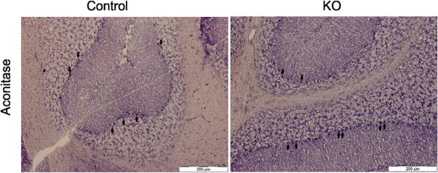
Figure 13.
In situ aconitase activity stain. An in situ histochemical staining for aconitase, a Fe–S containing protein, activity revealed no difference between PCs of control and KO mice at 6 months of age because all of the PCs also stained positively. PCs were identified by visual inspection according to location and shape of the neurons. Arrows point at examples of stained PCs. Scale bar, 200 μm.

