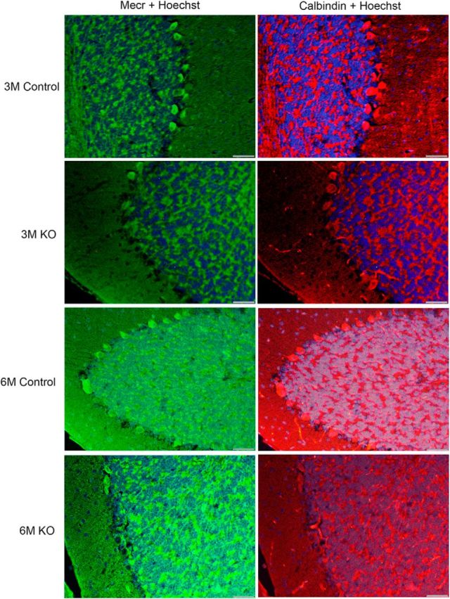
Figure 3.

IF studies of MECR and calbindin distribution in cerebella of control and PC-specific Mecr KO mice aged between 3 and 6 months. Shown are cerebellar sections probed with two different primary antibodies and corresponding secondary antibodies carrying green or red fluorescent tags identifying MECR and calbindin proteins, respectively. A Hoechst 33342 stain was used to label cellular nuclei. Scale bar, 40 μm.
