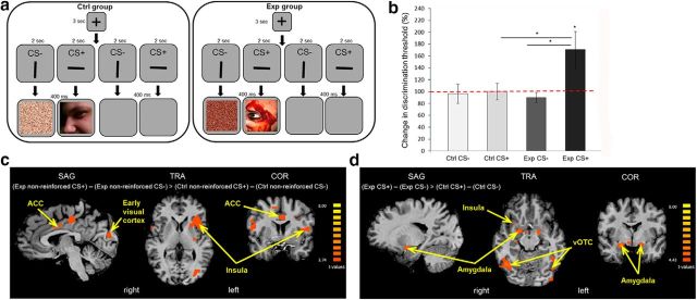
Figure 4.
Brain activity is modulated by visual aversive learning (Experiment 4). a, A schematic representation of one trial from the conditioning session of the fMRI experiment (Experiment 4). The paradigm was similar to the one described in Figure 3a, except for a partial conditioning of 50%: half of the trials were reinforced trials (CS+ stripe paired with image or CS− stripe paired with scrambled image), and the other half were nonreinforced trials (CS+ or CS− stripes paired with blank screens). b, Discrimination thresholds for orientation were tested before and after the conditioning session (experimental group, n = 30; control group, n = 29). No change in thresholds was measured in both groups for the CS− stripe, or in the control group for the CS+ stripe. The experimental group deteriorated (increase in threshold) in the CS+ orientation (paired with aversive images), with a CS × group interaction effect. c, A contrast of (experimental group nonreinforced CS+ trials − experimental group nonreinforced CS− trials) > (control group nonreinforced CS+ trials − control group nonreinforced CS− trials) showed higher activations in the ACC, insula, and early visual cortex in the experimental group (Exp) compared with the control group (Ctrl). Activations are shown in statistical q(FDR) < 0.05 thresholds. d, A contrast of (experimental group CS+ trials − experimental group CS− trials) > (control group CS+ trials − control group CS− trials) showed higher activations in the amygdala, insula, and vOTC in the Exp compared with the Ctrl. Activations are shown in statistical q(FDR) < 0.05 thresholds. *p < 0.05, see text for details regarding specific significance values.

