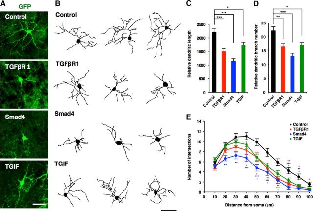
Figure 9.
TGIF-mediated canonical TGF-β signaling downregulates neuronal morphogenesis in vivo. A, Representative images of electroporated neurons, stained with anti-GFP antibodies. Mouse embryos were electroporated with plasmids expressing only GFP (control, green), or GFP together with TGFβR1, Smad4, and TGIF, by in utero electroporation at E14 and killed at P10. Scale bar, 50 μm. B, Representative tracing images of neurons electroporated with the constructs as in A. Scale bar, 50 μm. C, D, Quantification of total dendrite length (C) and dendrite branch numbers (D) in B. Number of cells analyzed: control, 18 neurons; TGFβR1, 12 neurons; Smad4, 13 neurons; TGIF, 16 neurons from three brains. E, Quantification of dendrite complexity by Sholl analysis of neurons in B. Data are presented as the mean ± SEM. *p < 0.05, **p < 0.01, ***p < 0.001 by one-way ANOVA, Tukey's post-test.

