Figure 2.
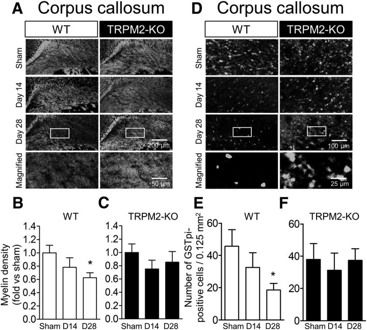
BCAS-induced white matter injury at day 28 was not observed in TRPM2-KO mice. A–C, Representative images of white matter in the corpus callosum by fluoromyelin staining (A) and the relative myelin density in WT (B) and TRPM2-KO mice (C). D–F, Representative images of immunostaining with GSTpi antibody in the corpus callosum (D) and the number of positive cells counted in WT (E) and TRPM2-KO mice (F). A, D, Bottom, Magnified images from the location marked by the boxed area of the above panels. *p < 0.05 vs WT sham. Values are mean ± SEM. B, C, n = 8–12; E, F, n = 6–9.

