
Figure 2.
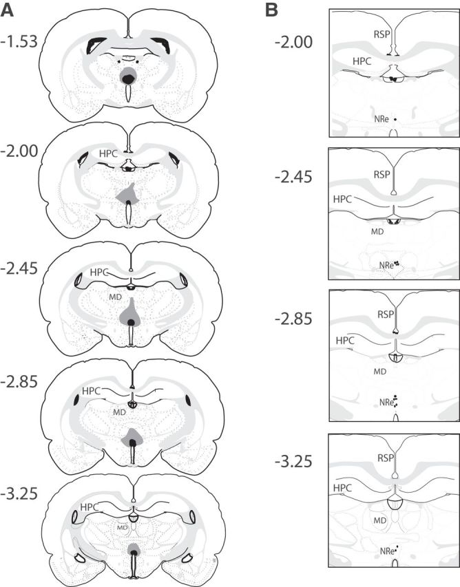
A, Diagrammatic reconstructions showing the cases with the largest (gray) and smallest (black) lesions in the ventral midline thalamic nuclei including the NRe and Rh. B, Diagrammatic representation of the induvial infusion sites in the NRe in each animal. The numbers correspond to the approximate position relative to bregma (Swanson, 1998). RSP, Retrosplenial cortex; MD, medial dorsal thalamic nuclei.