Figure 1.

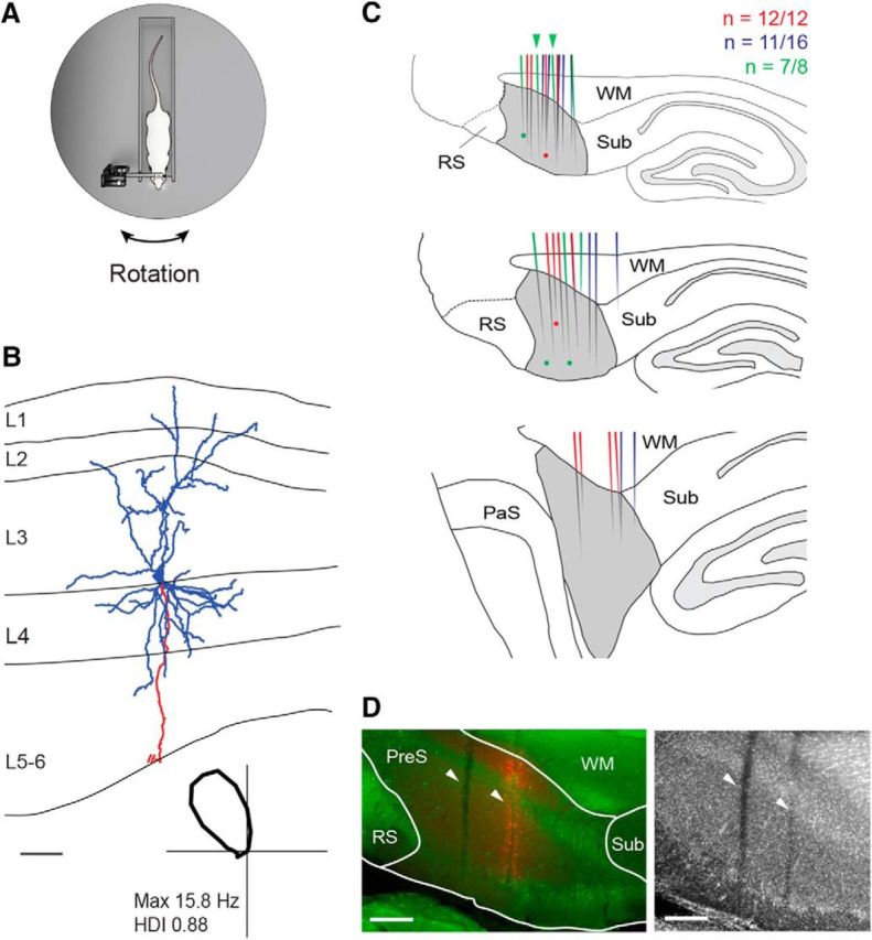
Juxtacellular recordings of presubicular HD neurons in head-fixed rats. A, Schematic representation of the Open recording configuration, consisting of a head-fixed rat on a rotating platform in the presence of a rich set of proximal and distal cues (see also Preston-Ferrer et al., 2016). B, Reconstruction of the dendritic (blue) and axonal morphology (red) of a layer 3 pyramidal HD cell recorded in a head-fixed rat. The axon is truncated for display purposes. The borders of the presubicular layers are indicated (L1–L6). Bottom, Polar plot showing the directional tuning for the representative reconstructed cell. Peak firing rate and HD index are indicated. Scale bar, 100 μm. C, Schematic outline of parasagittal sections through the rat brain at three mediolateral extents of the dorsal PreS (gray): medial (top), intermediate (middle), and lateral (bottom) (see Materials and Methods for details). The locations of reconstructed tracks through the PreS (color lines) and identified cells (color dots) are represented in different colors for three representative brains (the number of recovered tracks of penetration attempts are indicated). Green arrowheads indicate the two representative tracks shown in D. WM, White matter (angular bundle), Sub, subiculum, RS, retrosplenial cortex, PaS, parasubiculum. Scale bar, 500 μm. D, Left, Parasagittal section stained for calbindin (green) and neurobiotin (red) showing two electrode tracks (white arrowheads) through the dorsal PreS. In the anterior penetration, spillover of Neurobiotin was performed to aid anatomical recovery of the track (see details in Materials and Methods). Right, Same section stained for NeuN (the two tracks are indicated by the arrowheads).WM, White matter (angular bundle), Sub, subiculum, RS, retrosplenial cortex. Scale bars, 200 μm.
