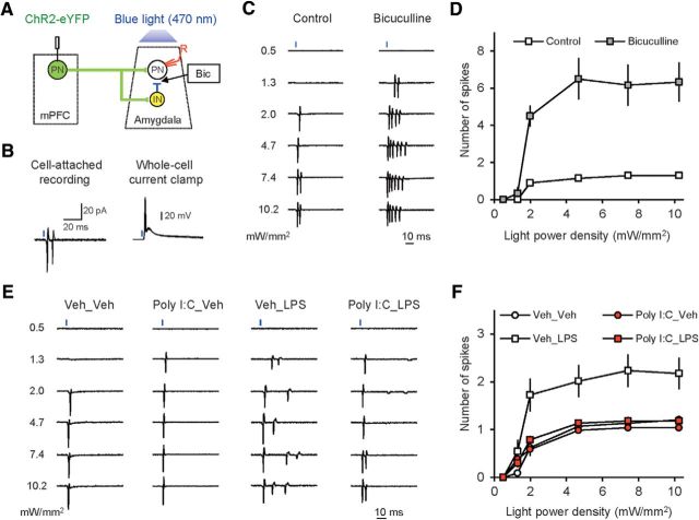
Figure 7.
Neuroinflammation is associated with the increased probability of synaptically driven spike firing in the mPFC–BLA pathway. A, Experimental design for recording of synaptically driven extracellular spikes in BLA PNs in response to photostimulation of mPFC fibers. B, Left, Superimposed synaptic responses (extracellular spikes) recorded in a BLA PN evoked by photostimulation of ChR2-expressing mPFC fibers in a cell-attached patch configuration. Right, Recordings under current-clamp conditions from the same neuron after establishing a whole-cell recording configuration. C, Examples of responses (extracellular spikes recorded in a cell-attached patch configuration) in BLA neurons to light pulses of increasing intensity under control conditions (left) and in the presence of the GABAA receptor antagonist bicuculline (30 μm, right). D, Number of spikes in response to presynaptic photostimulation was increased significantly in the presence of the GABAA receptor antagonist bicuculline (30 μm) in the external medium (control: n = 10 neurons from 3 mice; bicuculline: n = 6 neurons from 3 mice). E, Examples of synaptically driven spikes in BLA PNs in slices from different groups of mice. F, Summary input/output plots for synaptically driven spikes in BLA neurons in all experimental groups (Veh_Veh group: n = 22 neurons from 7 mice; Poly I:C_Veh: n = 13 neurons from 5 mice; Veh_LPS: n = 21 neurons from 5 mice; Poly I:C_LPS: n = 22 neurons from 7 mice).

