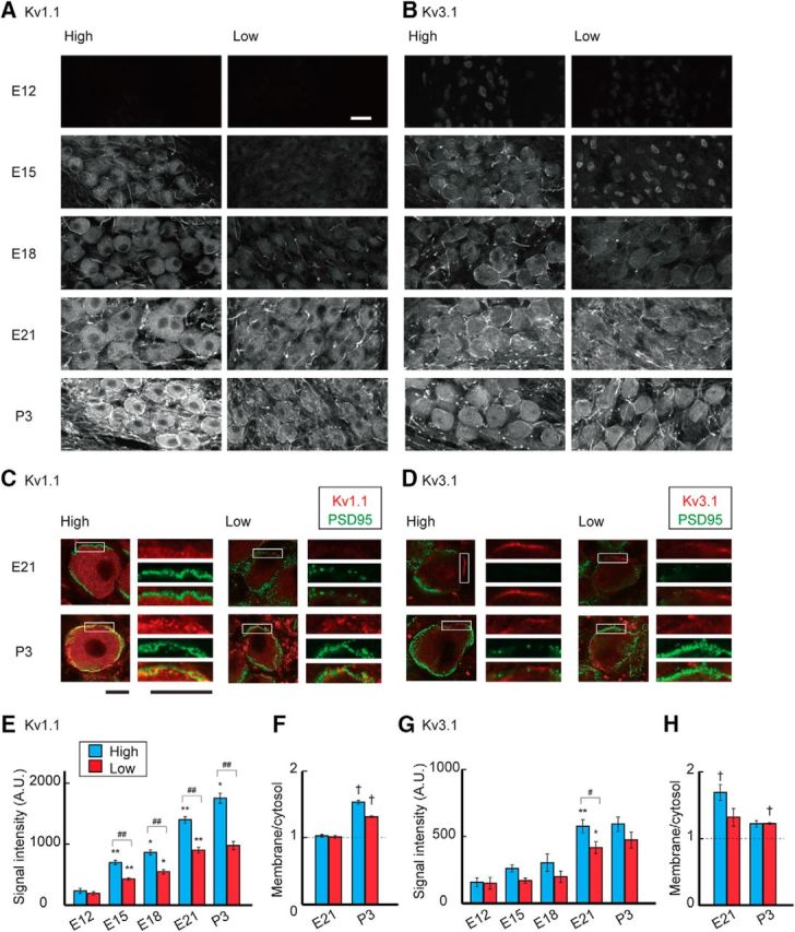
Figure 3.

Development of Kv1.1 and Kv3.1 expression. A, B, Immunosignals of Kv1.1 (A) and Kv3.1 (B) in high-CF (left) and low-CF (right) regions of the NM between E12 and P3. Immunosignals increased after E12, whereas the increase was greatly augmented and membranous signals appeared for Kv1.1 after hatch particularly in the high-CF region. Fibrous signals could be observed after E15 for both Kv1.1 and Kv3.1, suggesting that axonal targeting of these channels progresses during the embryonic period (Kuba et al., 2015). C, D, Double immunostaining of Kv channels (red) and PSD95 (green) at E21 (top) and P3 (bottom). Right, Boxes are magnified. C, Kv1.1. D, Kv3.1. PSD95 signals surrounded the soma and intermingled with membranous Kv signals at P3, confirming that the Kv signals are on the plasma membrane of postsynaptic NM neurons. E, G, Signal intensity of Kv1.1 (E) and Kv3.1 (G) measured at cytosolic regions in the soma (see Materials and Methods). F, H, Ratio of membranous and cytosolic signal intensities of Kv1.1 (F) and Kv3.1 (H) at E21 and P3 (see Materials and Methods). Measurements were made from 3–7 animals in each group. Scale bars: A, B, 20 μm; C, D, 10 μm. #p < 0.05 and ##p < 0.01 between tonotopic regions. *p < 0.05 and **p < 0.01 compared with the neighboring younger group. †p < 0.05 between cytosolic and membranous signals.
