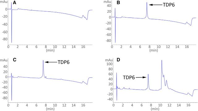
Figure 6.
TDP6 was retrieved and detected after 7 d incubation within osmotic minipumps implanted in conditional knock-out mice. A, Reverse-phase HPLC UV trace of aCSF (“blank/control” sample). B, HPLC UV trace of TDP6 at day before animal administration. C, HPLC UV trace of TDP6 at day 0. D, HPLC UV trace of TDP6 at day 7. All traces measured using 214 nm wavelength. Note: Additional peak in D was determined not to be a peptide degradation product. Area under the peptide peak was quantified in B–D and no significant change was observed.

