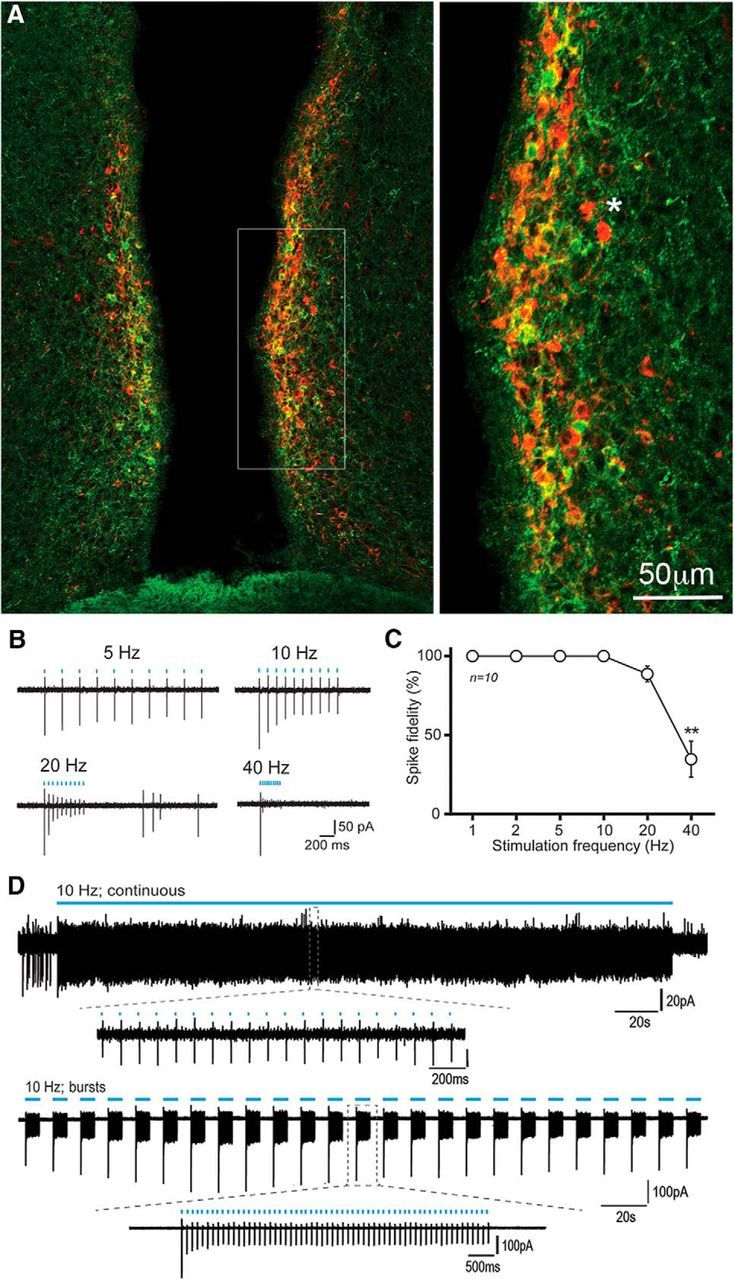
Figure 1.

ChR2-mCherry control of RP3VKISS neuron firing. A, Dual immunofluorescence showing kisspeptin (green), mCherry (red), and dual-labeled (yellow/orange) cells in the RP3V of an AAV-ChR2-mCherry injected Kiss1-IRES-Cre mouse. Inset (expanded to right), The relative intensities of the GFP and mCherry result in many cells being orange. An asterisk indicates two cells expressing only mCherry for reference. B, Example traces illustrating the response of an RP3VKISS neuron to trains of 10 blue-light pulses delivered at 5, 10, 20 and 40 Hz. C, Summary graph of spike fidelity as a function of stimulation frequency in 10 different RP3VKISS neurons. Spike fidelity significantly decreases at 40 Hz (**p < 0.01). D, Example traces illustrating the response of RP3VKISS neurons to a 5 min blue light stimulation train at 10 Hz (continuous) and to burst stimulation (60 pulses at 10 Hz every 12 s) delivered over 5 min (bursts). Insets, Individual spikes at expanded timescales.
