Figure 2.

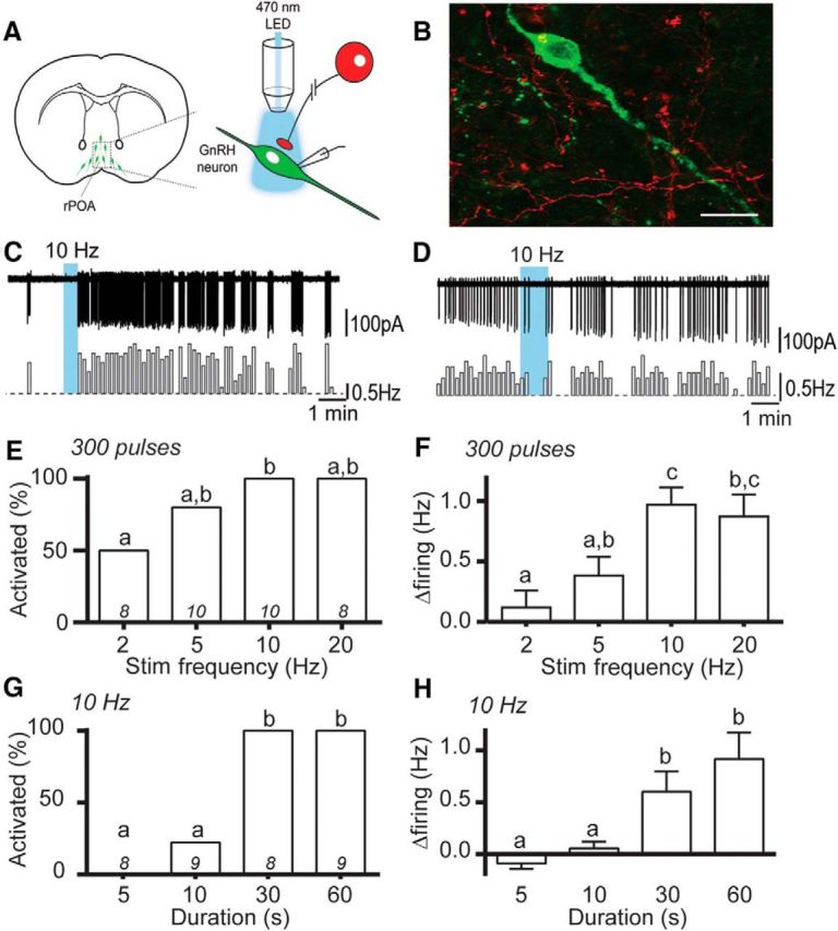
Optogenetic activation of RP3VKISS neuron axons excites GnRH neurons. A, Schematic representation of optogenetic brain slice experiment. B, Photomicrograph showing a GnRH-GFP neuron surrounded by RP3V origin mCherry-positive fibers opposing its cell body and primary dendrite (yellow overlap). C, D, Example traces and corresponding rate meters below (10 s bins) of GnRH neurons that increased its firing rate (C) or was unaffected (D) by 10 Hz blue-light stimulation of RP3VKISS neuron axons. E, F, Increasing stimulation frequency, with a constant number of pulses, increases the proportion of responding GnRH neurons (E) and the magnitude of their responses (F). G, H, Increasing the stimulus duration, at 10 Hz, increases the proportion of excited GnRH neurons (G) and the magnitude of their responses (H). Bars with differing letters are significantly (p < 0.05) different to each other.
