Figure 4.

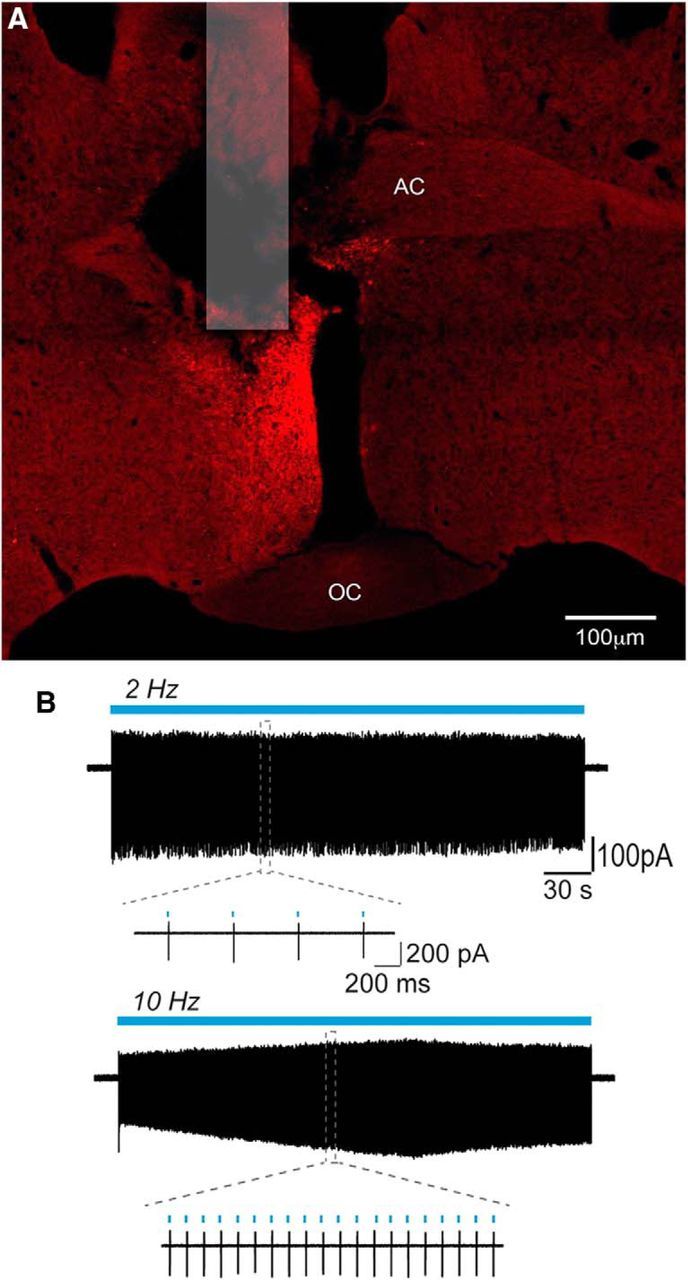
ChR2-mCherry control of RP3VGABA neuron firing. A, Photomicrograph showing location of fiber optic and expression of mCherry following unilateral injection of AAV-ChR2-mCherry into the RP3V of a Vgat-Cre mouse. AC, Anterior commissure; OC, optic chiasm. B, Example traces illustrating the response of an RP3VGABA neuron to 5 min continuous stimulation trains at 2 Hz (top trace) and at 10 Hz (bottom trace). Insets show individual spikes at expanded timescales.
