Figure 3.
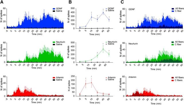
GDNF, neurturin, and artemin applied directly to the marrow cavity activates bone afferent neurons. A, Frequency histograms of the total number of spikes isolated from whole-nerve recordings before and after the application of GDNF, neurturin, or artemin (n = 5 animals each). Bin width, 30 s. Error bars indicate the SEM. B, Group data showing the number of spikes recorded (mean ± SEM) before and 5, 15, 30, 45, and 60 min after the application of GDNF, neurturin, artemin, or saline (n = 5 animals each). At each time-point, the number of spikes has been determined over five consecutive minutes and is represented as the mean ± SEM. Application of GDNF and neurturin resulted in an increase in whole-nerve activity, relative to saline, from the 15 and 30 min time-points, respectively (Bonferroni's post hoc test, *p < 0.05). The application of artemin resulted in a significant increase in whole-nerve activity, relative to saline, at the 5 and 15 min time-points, but not at the later time-points (Bonferroni's post hoc test, *p < 0.05). C, Frequency histograms showing the number of spikes with amplitudes consistent with Aδ- or C-fiber conduction velocities isolated from the same whole-nerve recordings. C-fiber spikes contributed more than Aδ-fiber spikes to the prolonged changes in activity after the application of GDNF, neurturin, and artemin, whereas Aδ-fiber spikes contributed to early activity induced by artemin.

