Figure 6.

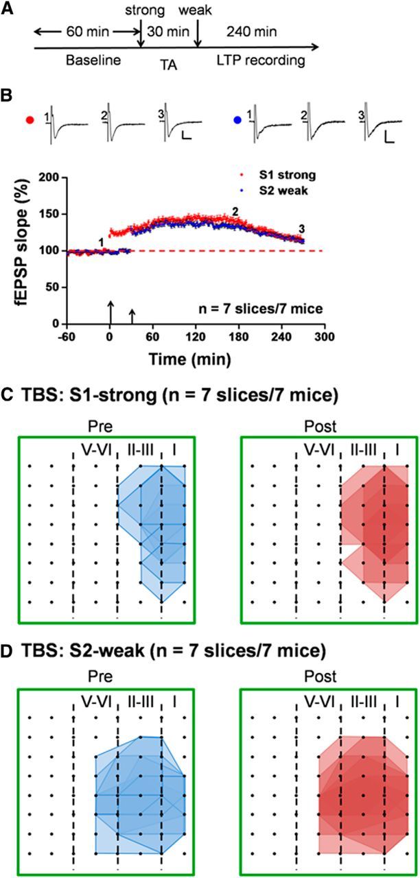
Loss of synaptic tagging in the ACC after tail amputation in adult mice. A, Schematic diagram of the recording procedure for this experiment. We applied the weak TBS protocol to the S2 site at 30 min after strong TBS of the S1 site in the ACC slices taken from tail-amputated mice (2 weeks experience). TA, Tail amputation. B, Pooled data of the fEPSP slope from all channels for both S1-strong and S2-weak inputs. Neither strong nor weak TBS can induce L-LTP (n = 7 slices/7 mice), indicating the loss of cortical synaptic tagging after amputation. Inset, Representative fEPSP traces taken at the time points indicated by numbers in the graph. Calibration: 100 μV, 10 ms. Large arrow indicates starting point of strong TBS application; small arrow marks the time point of weak TBS delivery. Error bars represent SEM. C, Polygonal diagrams of the channels that were activated in the baseline state (blue, pre) and at 4.5 h after strong TBS of the S1 site (red, post) in seven slices from seven tail-amputated mice. Black dots represent the 64 channels in the MED64. Vertical lines indicate the layers in the ACC slice. D, Pooled data of the spatial analysis similar as in C but for the weak TBS of the S2 site (n = 7 slices/7 mice). Tail amputation blocked strong TBS-induced network potentiation in the ACC.
