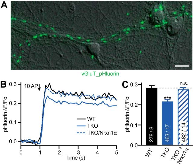
Figure 3.
Synaptic vesicle release is reduced in neurons lacking all α-neurexins. A, ΔF fluorescence image of vGlut_pHluorin (green) from a 100 AP stimulation, overlayed on DIC image of WT neurons. Scale bar, 20 μm. B, Exocytotic response of vGluT_pHluorin averaged across multiple synapses; comparison of WT, TKO, and TKO transfected with Nrxn1α (N given in corresponding bars in C). C, Summary of mean peak vGluT_pHluorin signals (ΔF/Fo) from conditions as in B. Data are mean ± SEM; n = ROIs/neurons (in bars), differences to WT are indicated. ***p < 0.001, n.s. = not significant, p = 0.454, by one-way ANOVA, F(2,1222) = 25.4.

