Figure 6.
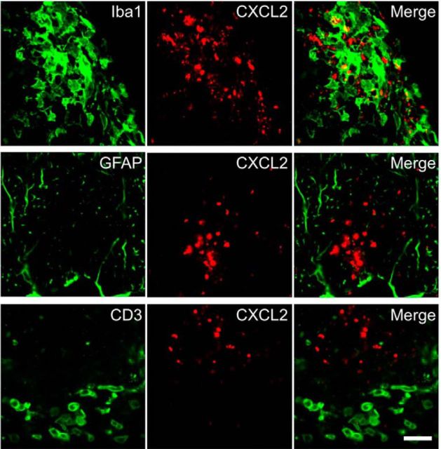
CXCL2 is primarily expressed by Iba1+ cells in the CNS. Representative lumbar spinal cord sections obtained from immunized mice at day 14 and stained with anti-Iba1 (top; macrophages/microglia), anti-GFAP (middle; astrocytes), anti-CD3 antibody (bottom; T cells), and anti-CXCL2 (center) antibodies are shown. Images of anti-CXCL2 antibody staining are shown in the center panels and merged images are shown in the right panels. Scale bar, 20 μm.

