
Figure 2.
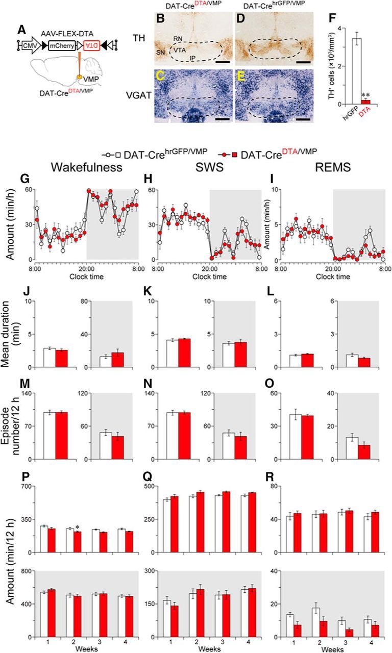
Ablation of the VMP dopaminergic neurons had little to no effect on wakefulness. A, DAT-Cre mice were injected with AAV-FLEX-DTA into the VMP. B–E, Brain sections stained against TH (B, D) or VGAT mRNA (C, E) from mice injected with AAV-FLEX-DTA (DAT-CreDTA/VMP mice; B, C) or AAV-FLEX-hrGFP (DAT-CrehrGFP/VMP mice; D, E) to confirm that the VMP dopaminergic neurons were selectively ablated in DAT-Cre mice. Dashed line delineates the area with loss of dopaminergic neurons in a DAT-CreDTA/VMP mouse. Scale bar, 500 μm. F, Number of neurons expressing TH in the VMP of mice injected with AAV. G–I, Time course of hourly wakefulness (G), SWS (H), and REMS (I) at 1 week after surgery. J–O, Ablation of the VMP dopaminergic neurons changed neither the mean duration (J–L) nor the number (M–O) of each episode. P–R, Total amount of wakefulness (P), SWS (Q), and REMS (R) for 12 h. White and gray background in each panel indicate light and dark periods, respectively. *p < 0.05, **p < 0.01 compared with DAT-CrehrGFP/VMP mice. Data are presented as the mean ± SEM (n = 6). RN, Red nucleus; SN, substantia nigra; IP, interpeduncular nucleus.