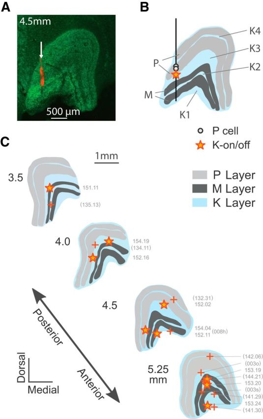
Figure 2.

Anatomical reconstruction of K-on/off cell positions. A, coronal section through the LGN (4.5 mm anterior to intra-aural line) stained with NeuroTrace (green) and DiI (red), showing the layers of the LGN and electrode track (arrow). B, Reconstructed positions of two P cells and one K-on/off cell recorded in this track. C, Reconstructed positions of K-on/off cells (star symbols) shown relative to representative LGN profiles. The positions of the other K-on/off cells were estimated from receptive field location and eye dominance relative to other cells recorded on the same track (+ symbols).
