Figure 1.
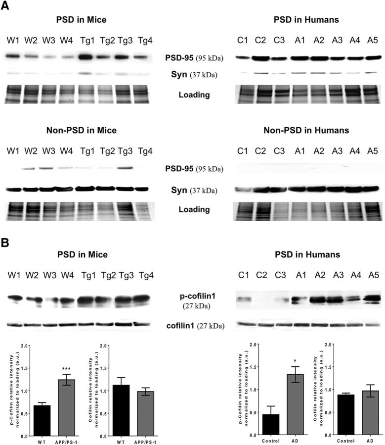
Cortical samples from APP/PS1 mice and AD cases have elevated p-cof1. A, Western blot images of PSD-enriched (top) and non-PSD fraction (bottom) of cortical synaptosomes of APP/PS1 mice (left) and humans (right) showing the enrichment of presynaptic and postsynaptic marker in each fraction. B, Western blot images (top) along with associated quantification (bottom) showing elevated phospho-cofilin1 in APP/PS1 mice (left) compared with their nontransgenic littermates (Western blots n = 4; p-cof1 Mann–Whitney U = 6.0 (72,138) p = 0.0011; cof1 Mann–Whitney U = 43.0 (102,88) p = 0.9025) and in AD cases (right) compared with healthy controls (WB, n = 4; p-cof1 Mann–Whitney U = 0.0 (6,30) p = 0.0357; cof1 Mann–Whitney U = 7.0 (13,23) p = 1.000). *p < 0.05. ***p < 0.001. For case details, see Table 1.

