Figure 7.
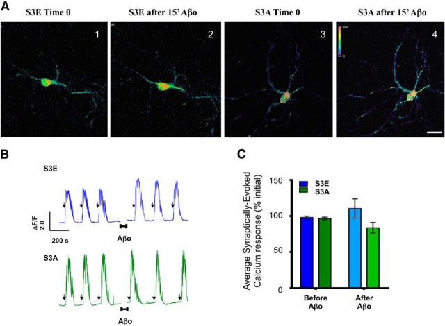
cof1 phospho-mutants prevent impairment of synaptic calcium responses induced by Aβo. A, Representative images of primary cortical neurons (DIV 14) transfected with GCaMP6fast (an indicator of synaptically driven calcium responses) and inactive cof1 phosphomimetic (S3E) or the nonphosphorylatable cof1 (S3A) during 15 s pulses of B4AP before and after 15 min incubation with Aβo. B, Representative ΔF/F traces of GCaMP6fast fluorescence as an indicator of synaptically driven calcium responses induced by 15 s pulses of B4AP (arrows) in neurons expressing the inactive cof1 phosphomimetic (top, S3E, blue), or the nonphosphorylatable cof1 (bottom, S3A, green), before and after 15 min incubation (indicated by the horizontal bars and break in the traces) with Aβo (100 nm). C, Quantification of the B4AP-evoked synaptic calcium responses from B, before (left bars, dark) and after (right bars, light) 15 min exposure to Aβo in neurons expressing the inactive cof1 phosphomimetic (S3E, blue) or the nonphosphorylatable cof1 (S3A, green). Both cof1 mutants block the Aβo-induced synaptic impairment (two-way ANOVA: time, F(1,17) = 0.0006, p = 0.983; treatment, F(1,17) = 3.257, p = 0.0889; interaction, F(1,17) = 0.8935, p = 0.142). Error bars indicate mean ± SEM. n = 9 or 10 neurons, 6 or 7 neuronal cultures per group.

