Figure 8.
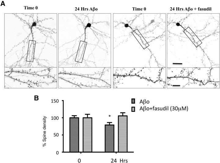
Fasudil treatment reverses Aβo-induced synaptotoxicity. A, Representative images of DIV 14–15 neurons expressing actin-GFP before (t 0) and after incubation 24 h with Aβo or fasudil + Aβo. Bottom panels, Magnified detail of the selected area in top panels, respectively. Scale bars: Top, 30 μm; Bottom, 10 μm. B, Relative quantification of spine density in neurons expressing actin-GFP before and after incubation 24 h with Aβo or fasudil + Aβo. A paired t test showed that 24 h exposition to Aβo caused decrease in the spine density that was blocked by fasudil (t values t(10) = 8.931, p < 0.0001). Error bars indicate mean ± SEM. n = 11 neurons, 3 cultures per group. *p < 0.05.

