
Figure 9.
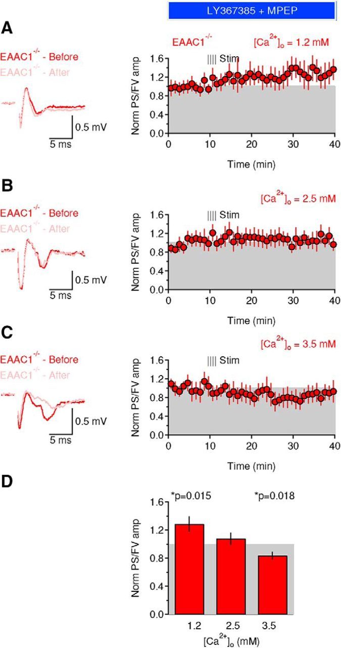
Blocking mGluRI activation rescues the Ca2+ dependence of long-term plasticity in EAAC1−/− mice. A, Left, Extracellular recordings performed in the presence of the mGluRI blockers LY367385 (50 μm) and MPEP (10 μm) obtained 5 min before (red trace) and 30 min after applying an HFS protocol (100 Hz, 1 s) to the DLS of EAAC1−/− mice (pink trace). The recordings were obtained in the presence of extracellular solutions containing [Ca2+] = 1.2 mm. Each trace represents the average of 20 consecutive sweeps. The shaded area represents the SEM. Right, Time course of baseline-normalized field recordings. Each symbol represents the average of three consecutive time points. The notation “Norm PS/FV” on the y-axis refers to the amplitude ratio of the population spike and fiber volley normalized by the one measured before applying the HFS protocol. B, As in A but in the presence of extracellular solutions containing [Ca2+] = 2.5 mm. C, As in A but in the presence of extracellular solutions containing [Ca2+] = 3.5 mm. D, Summarized effect of HFS on the PS/FV ratio in EAAC1−/− mice in the presence of mGluRI blockers and at three different extracellular [Ca2+] [1.2 mm (n = 22), *p = 0.015; 2.5 mm (n = 12), p = 0.36; 3.5 mm (n = 8), *p = 0.018].