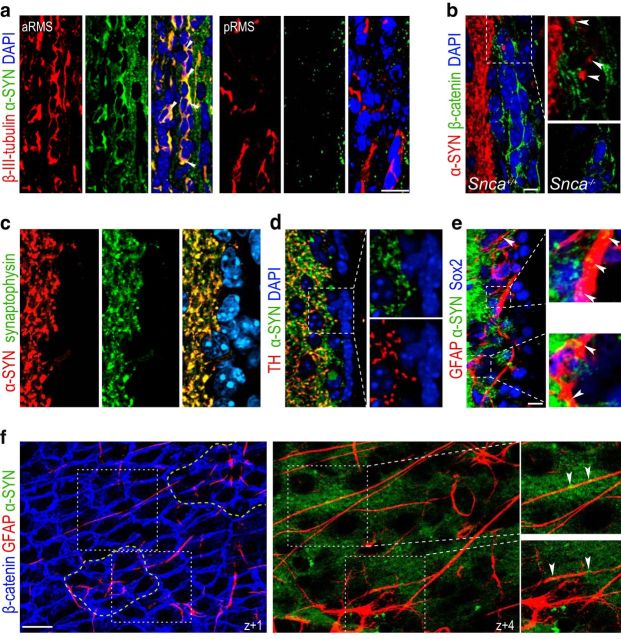
Figure 2.
α-SYN is present in fibers supplying the SEZ. a, Immunofluorescent detection of α-SYN (green) and β-III-tubulin (red) in coronal sections through the anterior (a) and posterior (p) half of the RMS. White arrowheads point at doubly positive cells present only in the aRMS. DAPI was used as a counterstain. b, Immunofluorescent detection of α-SYN (red) and β-catenin (green) to label cell membranes in the SEZ in Snca+/+ and Snca−/− mice as a specificity control. White arrowheads point at puncta within the SEZ region. c, Synaptic distribution of α-SYN (red) observed by colocalization with synaptophysin+ (green). d, Synaptic colocalization of α-SYN (green) with TH+ (red). There is characteristic punctate staining of nerve terminals and the lack of staining in SEZ cells in the high-power micrographs (right) of the area indicated by the square. e, Low-power confocal micrographs showing the immunofluorescent detection of α-SYN (green), GFAP (red), and Sox2 (blue) and high-power images of the areas indicated by the white squares. α-SYN+ terminals are closely adjacent to GFAP+Sox2+ cells (indicated by white arrowheads). f, Serial confocal sections through a SEZ whole mount at different z levels from the ventricular surface (z + 0) show the staining for β-catenin (blue), GFAP (red), and α-SYN (green). Dotted yellow lines indicate two pinwheels. Different GFAP+ B1 cells traced in the z-axis from the ventricular surface with α-SYN+ puncta closely apposed (white arrowheads) as shown at a higher magnification of the dotted white lined square. DAPI (blue) was used for nuclear staining. Scale bars: a, 20 μm; b–f, 10 μm.

