Figure 4.
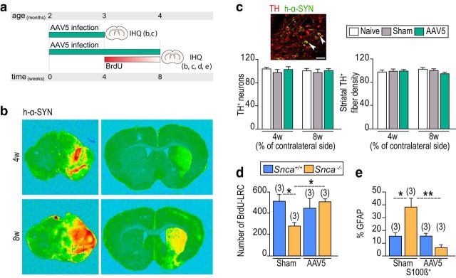
α-SYN present in DAergic nigral synaptic terminals prevents differentiation of NSCs into non-neurogenic astrocytes. a, Experimental protocol for AAV5-CBA-h-α-Syn SNpc infection and subsequent analysis. b, Detection of human α-SYN expression in the hemispheres ipsilateral and contralateral to the infection at the levels of the mesencephalon and striatum 4 and 8 weeks after the surgery. c, Representative immunofluorescent detection of TH (red) and α-SYN (green) in the SNpc ipsilateral to the viral injection. Quantification of the number of TH+ neurons and the density of TH+ fibers in the striatum in naive, sham, and AAV5-CBA-h-α-SYN mice, evaluated 4 and 8 weeks after the adenovirus injection. d, Quantification of BrdU-LRCs in the SEZ of sham-operated and virus-infected Snca+/+ and Snca−/− mice. e, Histogram showing the proportions of GFAP+ subependymal cells that are also S100β+ in sham or infected Snca+/+ and Snca−/− mice 8 weeks after the surgery. *p < 0.05 (two-way ANOVA). **p < 0.01 (two-way ANOVA). Scale bar: c, 20 μm.

