Figure 2.
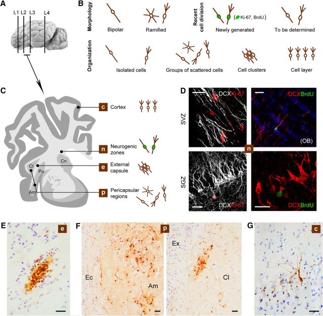
DCX+ cells in the adult sheep brain. A, C, Representative levels of the brain showing different locations of the DCX+ cells. B, Main types of DCX+ cells encountered in our analysis (DCX+ “objects”) classified according to their morphology, spatial organization, and cell division history (newly born vs non-newly generated). D, Newly generated neuroblasts in the SVZ and dentate gyrus (SGZ); in both neurogenic sites, DCX+ cells are intermingled with several nuclei immunoreactive for Ki-67 antigen (rarely double stained in the SVZ due to different expression time course of the markers); BrdU injected 60 d before the animals were killed is detectable in DCX+ neuroblasts of the olfactory bulb (OB) and in hippocampal granule cells. E–G, Representative photographs of the DCX+ cells/cell populations at the different locations showed in C. E, Clusters of DCX+ cells in the external capsule (Ec). F, Scattered DCX+ cells in the amygdala (Amy) and claustrum (Cl); Ex, capsula extrema. G, Layer II cortical neurons. Scale bars: 30 μm; D (bottom right), 20 μm.

