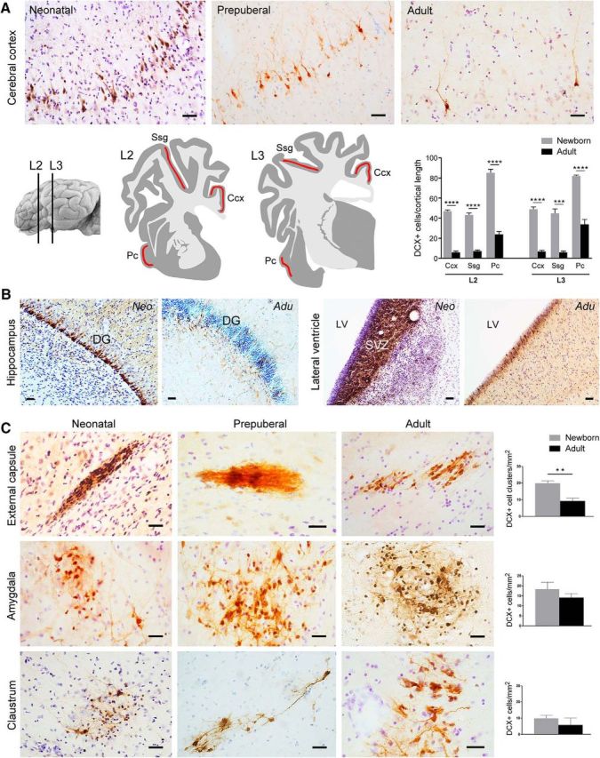
Figure 6.

DCX+ cells in the sheep brain at different ages. A, Evident reduction of the amount of DCX+ neurons in the cortical layer II with increasing age is clearly visible after qualitative analysis (top). Quantitative evaluation of DCX+ cell linear density (number of DCX+ neurons in layer II/cortical tract length; bottom) in three cortical regions (red areas) at two brain levels of the newborn and adult sheep. Pc, Piriform cortex; Ssg, suprasylvian gyrus; Ccx, cingulate cortex (***p = 0,0001, ****p < 0,0001). B, Striking reduction of DCX+ cell populations is clearly evident in the dentate gyrus (DG; note the dilution of the DCX+ cell layer) and SVZ (note the reduction in thickness of the DCX+ germinal layer) neurogenic sites of neonatal and adult sheep. C, Occurrence, morphology, distribution, and amount (quantifications on the right; see also Tables 3, 4, 5) of DCX+ cells in the sheep capsular/pericapsular regions do not vary significantly at different ages (apart from a slight reduction observed in the external capsule; see text). Scale bars, 30 μm (**p= 0,001).
