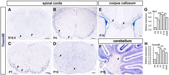
Figure 1.
Developmental expression pattern of Tmem98 in the CNS. A–D, In situ hybridization for Tmem98 was performed on the sections of spinal cord from E18.5 (A), P4 (B), P7 (C), and P15 (D) wild-type mice. E, F, Sections from P15 corpus callosum (E) and cerebellum (F) of wild-type mice were subjected to ISH with Tmem98 riboprobes. G, H, Transcriptional level of Tmem98 and Myrf mRNA at different developmental stages was quantified by RT-qPCR. Representative Tmem98-positive cells are indicated by arrows. *p < 0.05, **p < 0.01, ***p < 0.001. Scale bars, 100 μm.

