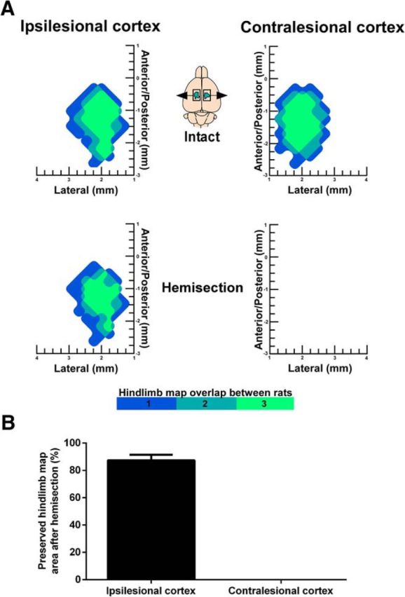
Figure 3.

Ipsilesional hindlimb motor maps are preserved 1 h after hemisection. A, Surface plots showing the frequency distribution of hindlimb motor maps derived with ICMS in the intact state and 1 h after hemisection (n = 3 rats). After hemisection, hindlimb motor maps were abolished in the contralesional cortex and preserved in the ipsilesional cortex. B, Quantification of hindlimb motor map area after hemisection relative to the intact state (%). Data are plotted as group mean ± SD.
