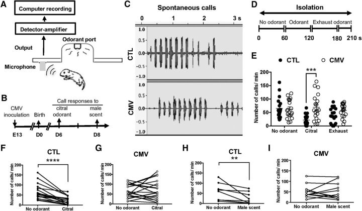
Figure 2.
Impact of CMV congenital infection on neonate olfaction. A, Emission and quantitation of ultrasonic vocalizations. The recording of ultrasonic calls began 30 s after placing the pups in the test chamber of the olfactometer. Ultrasonic vocalizations were detected using an ultrasonic microphone connected to a bat detector that converts ultrasonic sounds to the audible frequency range. B, Timetable of the experiments. Mice were infected in utero with CMV or received PBS at E13. They were analyzed using olfactometers at 6 and 8 d after birth. C, Typical wave traces of spontaneous call series from preweaning 6-day-old pups after congenital CMV infection. CTL mice were inoculated with PBS only. D, Experimental paradigm. Ultrasonic emission responses were recorded during the first period without odorant (1 min), followed by a period of odorant exposure (1 min) and finally the last period of exhaust odorant (1 min and 30 s). E–G, Emission of ultrasonic calls for citral odorant on day 6 after birth (n = 18 CTL, n = 19 CMV). H, I, Emission of ultrasonic calls for male scent odorant on day 8 after birth (n = 8 CTL, n = 11 CMV). p-values were calculated by Mann–Whitney test (E) or Wilcoxon matched-pairs signed–rank test (H, I). **p < 0.01, ***p < 0.001, ****p < 0.0001; mean ± SEM in E.

