Figure 5.

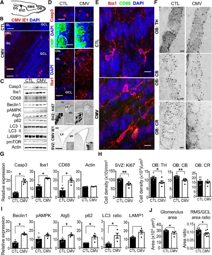
Impact of CMV congenital infection on the postnatal olfactory system at W3. A, Sagittal section of a murine brain showing the neurogenic dentate gyrus (DG) of the hippocampus, the neurogenic SVZ, the RMS, and the OB. Neuroblasts born in the neonatal and postnatal SVZ migrate via the RMS until the OB, where they differentiate into GCL or GL interneurons. B, D–F, Representative staining of coronal SVZ (D, bottom) and OB slices with DAPI (B, D, E) murine CMV IE1 (B, D, bottom), cleaved caspase 3 (Casp3, D, top), OMP expressed by OSN (D, middle), Iba (D, middle; E), CD68 expressed by activated microglia and macrophages (E), Ki67 (D), TH (F), CB (F), and CR (F), antibodies showing CMV+, apoptotic Casp3+, OSN, macrophages, microglia, Ki67+ neural progenitor cells, TH+, CB+ and CR+ cells in CTL and congenital CMV-infected mice at W3 after birth. C, G–I, Screening of the OB proteins from congenital CMV-infected mice at W3 for autophagy induction (C, I), cell apoptosis (C, G), and microglial reaction (C, G). Lysates were extracted from the OB of CTL and congenital CMV-infected mice at W3 and analyzed by immunoblot (C) using antibodies to detect Casp3, Iba1, CD68, Beclin1, phospho-AMPK, Atg5, p62, LC3 I/II, LAMP, phospho-mTOR, and actin (three mice each). The levels of Casp3, Iba, CD68, Beclin1, pAMPK, Atg5, p62, LC3 II/ LC3 I, LAMP, pmTOR, and actin were quantified (G,I) by band intensity with Fiji software. H, J, Ki67+, TH+, CB+, and CR+ cell densities in the SVZ, GL, glomerulus (glom), and GCL at W3 following congenital CMV inoculation. All mice are W3-old males injected at E13 with PBS (CTL) or CMV. For immunoblot analysis (G, I), n = 4 mice per group. For cell density analysis (H) and area ratio (J), n = 4–6 mice per group. For glom size (J), n = 409 glom from 4 CTL, n = 352 glom from 4 CMV. Results are shown as mean ± SEM. p-values were calculated by Mann–Whitney test. *p < 0.05, **p < 0.01. Scale bars: 100 μm in D, bottom; 50 μm in B, D, middle, and F; 25 μm in D, bottom; and 5 μm in D, top, and E.
