Figure 2.
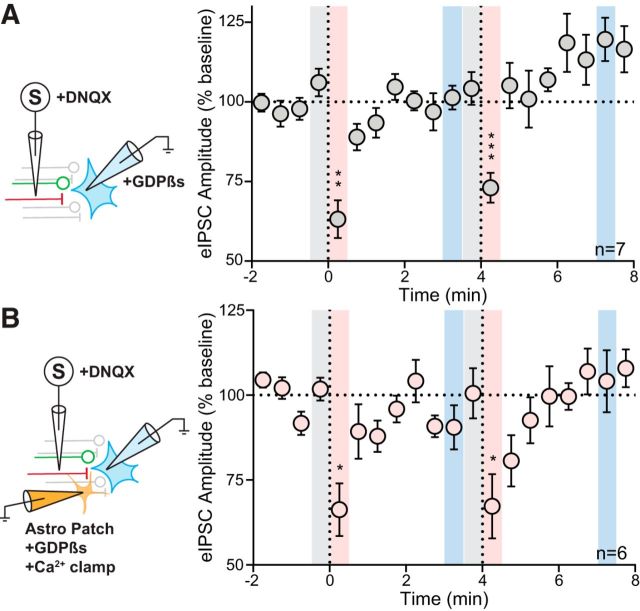
No role for astrocytic or postsynaptic GPCR signaling in LDSI. A, Left, Schematic showing postsynaptic loading of GDPβs. Right, Time course summary of 7 cells from 6 animals (4 male, 2 female) showing rapid recovery from DSI in neurons with impaired GPCR signaling (PD1: F(2,6) = 22.89, p = 0.0001, PD2: F(2,6) = 23.87, p = 0.0012). B, Left, Schematic showing astrocytes patched and filled with an internal solution that clamps Ca2+ transients and prevents GPCR activation. Following astrocytic loading, an adjacent neuron is patched and eIPSCs are elicited. Right, Time course summary of 6 cells from 5 animals (4 male, 1 female) showing that PNCs within the field of a patched astrocyte exhibit rapid recovery from DSI (PD1: F(2,5) = 13.61, p = 0.0040, PD2: F(2,5) = 5.23, p = 0.028). Post hoc versus baseline p-values are shown as follows: *p < 0.05, **p < 0.01, ***p < 0.001. Data are shown as mean ± SEM.

