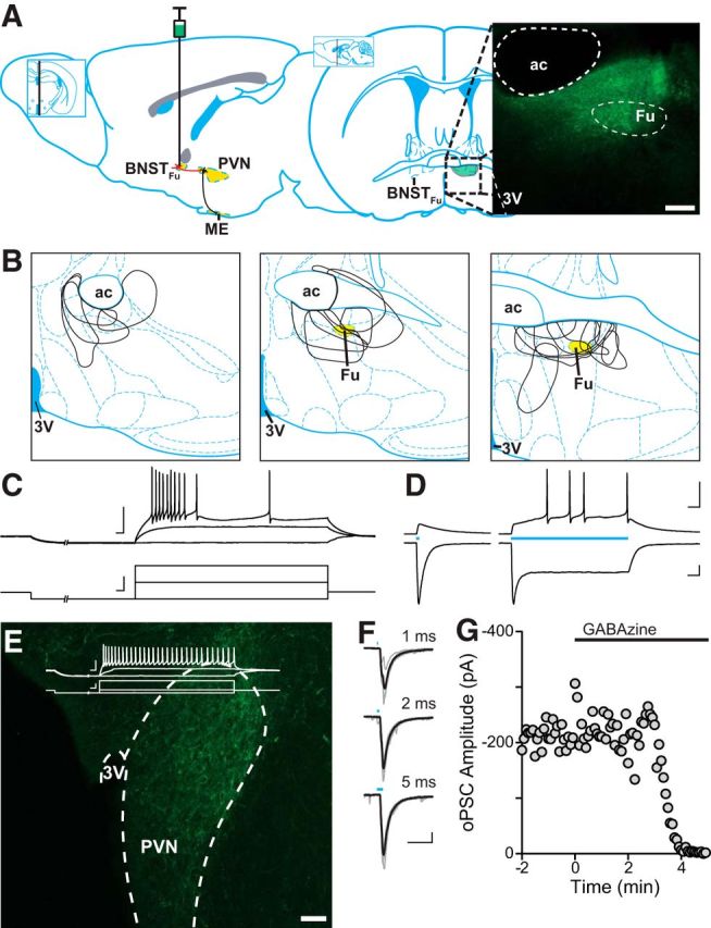
Figure 4.

Optically evoked GABA release in the PVN of vGAT-ChR2 BNSTFu mice. A, Left, Sagittal illustration denoting the projection from the BNSTFu to the PVN, targeted for stereotaxic viral injection. Right, Coronal map through the level of the BNST, indicating the location of the BNSTFu. Inset, Confocal image (10× magnification) showing eYFP expression in the BNSTFu of a vGAT-IRES-Cre mouse. B, Injection maps outlining BNSTFu targeting. C, eYFP-positive neurons from the BNSTFu were patched and characterized by their membrane voltage response before being exposed to varying durations of 473 nm light (D). BNSTFu neurons exhibited a characteristic steady-state current in response to a sustained exposure to blue light. E, Confocal image (20× magnification) depicting ChR2-eYFP-positive BNSTFu afferent fibers innervating the PVN. Inset, Representative trace showing a PNC membrane voltage response. F, Synaptic currents evoked by 473 nm light of varying durations. A 2 ms pulse was found to elicit synchronous release and was used for all other experiments. G, oPSCs were completely inhibited in a single cell by the application of the selective GABAA antagonist GABAzine (100 μm). Scale bars: A, E, 100 μm; C–F, 50 mV/pA, 20 ms.
