Figure 7.
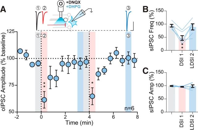
mGluR5 coactivation curtails optically induced LDSI. A, Top, Representative traces during DHPG application (50 μm). Inset, Experimental setup. Bottom, Summary time course of 6 cells from 6 mice (2 male, 4 female) showing postsynaptic depolarizations in DHPG repeatedly elicit DSI, but not LDSI (PD1: F(2,5) = 19.59, p = 0.0010, PD2: F(2,5) = 16.63, p = 0.0038). B, C, Summary graphs of a phasic suppression of sIPSC frequency (F(2,5) = 10.30, p = 0.0037; B), whereas sIPSC amplitude is unaffected by postsynaptic depolarization (F(2,5) = 0.29, p = 0.67; C). Scale bars, 100 pA, 20 ms. Post hoc versus baseline p-values are shown as follows: *p < 0.05, **p < 0.01. Data are shown as mean ± SEM.

