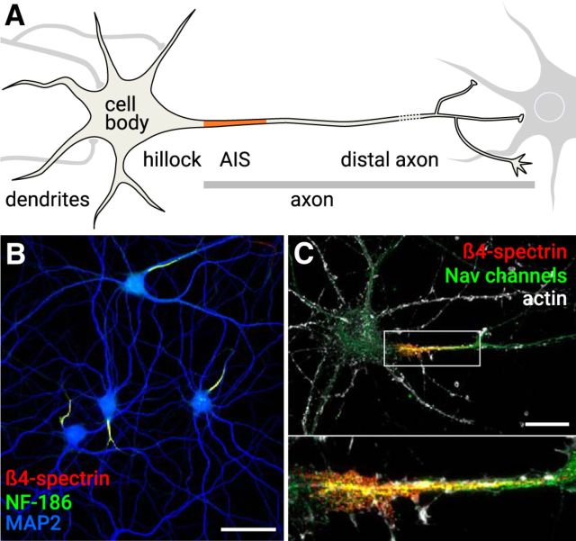
Figure 1.
The AIS. A, A typical neuron receives input on the cell body and dendrites (left). The hillock leads to the axon, which contains the AIS (orange). The distal axon contacts downstream neurons (right). B, Hippocampal neurons after 22 d in culture labeled for the AIS components NF-186 (green) and β4-spectrin (red). The somatodendritic compartment is labeled using an anti-MAP2 antibody (blue). Scale bar, 50 μm. C, Hippocampal neuron after 14 d in culture labeled for actin (gray), β4-spectrin (red), and Nav channels (green). Bottom, The zoomed image represents the AIS. Scale bar, 20 μm.

