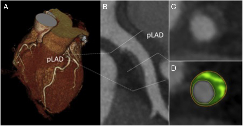
Figure 2.
Coronary computed tomography angiography (CCTA) (A) 3d volume rendering and (B) curved planar reformat through a proximal left anterior descending plaque. Plaque volume is quantified on the (C) short axis slice through the artery, with the (D) attenuation of plaque voxels categorized as noncalcified or calcified.

