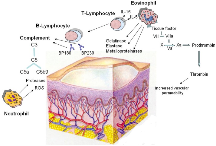
Figure 2.
Pathomechanisms of the blister formation in bullous pemphigoid including the involvement of coagulation activation. The dermo-epidermal detachment is due to the interaction of autoantibodies with two hemidesmosomal antigens (BP180 and BP230), followed by complement activation and leukocyte infiltration. Autoreactive T lymphocytes cooperate with B lymphocytes in the autoantibody production; in addition, they release cytokines, most notably IL-5 and IL-16, and other soluble factors responsible for the recruitment and activation of eosinophils. In lesional skin, eosinophils produce and release metalloproteinases, elastase, and gelatinase which contribute to tissue damage. Moreover, eosinophils strongly express tissue factor, which is the main initiator of the coagulation cascade (factors VII, X, VIII, V, and prothrombin) leading to generation of thrombin. This last increases the permeability of blood vessels, amplifying the inflammatory network. Neutrophils contribute in the pathogenesis of bullous pemphigoid by releasing reactive oxygen species (ROS) and proteases. Finally, complement is activated upon binding of the pathogenic autoantibodies to their autoantigens.

