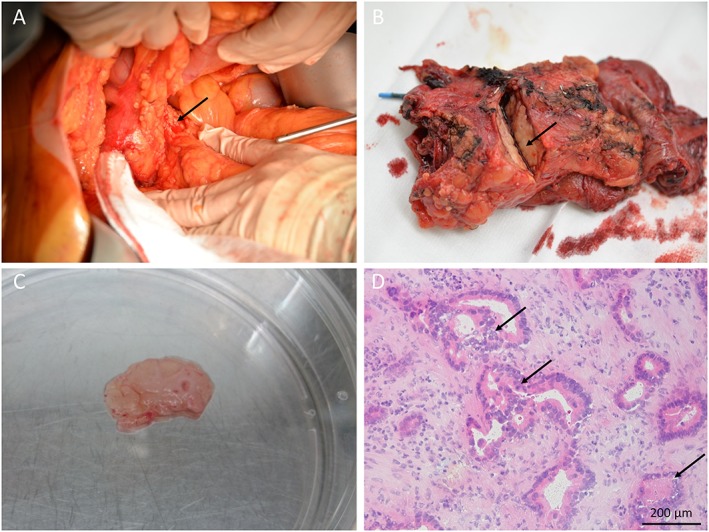
Figure 4.

Pancreatic tumour biopsy. (A) The pancreas head (arrow) is visible during pancreatic surgery. (B) The pancreas specimen is incised by the pathologist revealing the pancreatic tumour (arrow). (C) Tumour containing tissue slice. (D) Frozen section of the tissue slice confirming the presence of adenocarcinoma (arrows) (haematoxylin/eosin staining).
