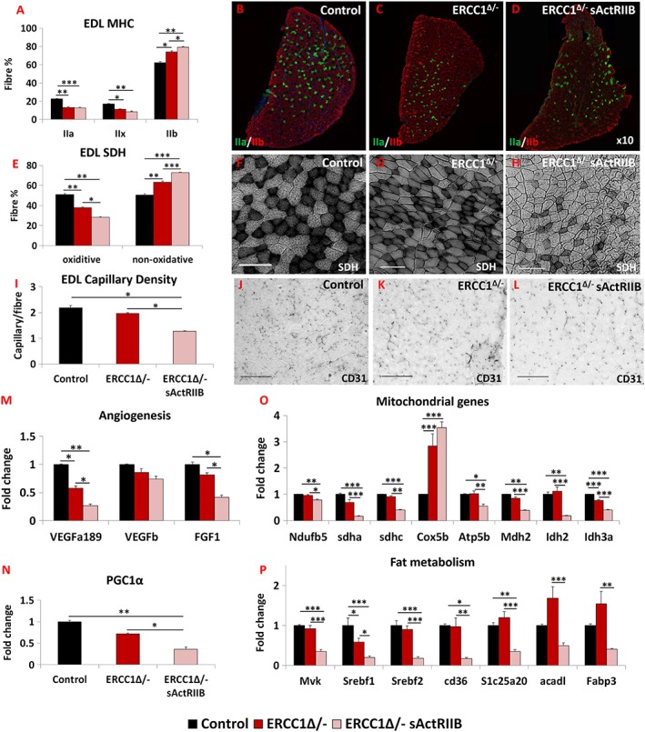
Figure 3.

sActRIIB induces fast and glycolytic transformation of Ercc1 Δ/− muscle. (A) MHC profile of EDL muscle. (B–D) EDL MHCIIA/IIB fibre distribution in the three cohorts, controls, Ercc1 Δ/−, and Ercc1 Δ/− treated with sActRIIB. (E) SDH‐positive and ‐negative fibre profile of EDL muscle. (F–H) SDH stain in the three cohorts. (I) Quantification of EDL capillary density. (J–L) Identification of EDL capillaries with CD‐31 in the three cohorts. Quantitative PCR profiling of (M) angiogenic genes, (N) PGC1α, (O) mitochondrial genes, and (P) regulators of fat metabolism. n = 8 for all cohorts. Scale for SDH 100 μm and CD31 50 μm. One‐way analysis of variance followed by Bonferroni's multiple comparison tests used in all data sets except (E) where non‐parametric Kruskal–Wallis test followed by the Dunn's multiple comparison was used. *P < 0.05, **P < 0.01, ***P < 0.001. EDL, extensor digitorum longus; MHC, myosin heavy chain; SDH, succinate dehydrogenase; sActRIIB, soluble activin receptor type IIB.
