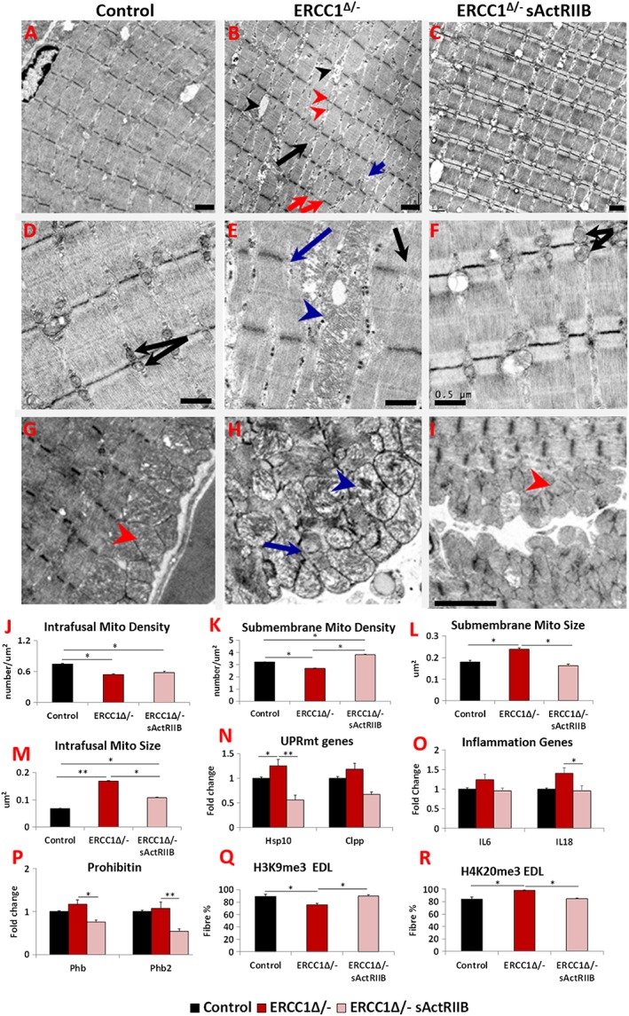
Figure 4.

sActRIIB prevents Ercc1 Δ/− muscle ultrastructural abnormalities and supports normal levels of expression of key stress indicators. All Electron microscopy (EM) longitudinal image and quantitative measurements are from the bicep muscle. (A) Low‐power image of control muscle. (B) Low‐power image of Ercc1 Δ/− muscle. Note large spaces (black arrowheads), non‐uniform sarcomere width (red arrows), dilated sarcomeric mitochondria (red arrowheads), split sarcomere (black arrow), and disrupted M‐Line (blue arrow). (C) Low‐power image of sActRIIB‐treated Ercc1 Δ/− muscle. (D) Higher magnification of sarcomeric region of control muscle showing uniformly sized mitochondria (black arrows). (E) Enlarged mitochondria in sarcomeric region of Ercc1 Δ/− muscle (blue arrowhead) and absent (blue arrow) or faint Z‐line (black arrow). (F) Higher magnification of sarcomeric region of treated Ercc1 Δ/− mice showing smaller sarcomeric mitochondria (black arrows). (G) Sarcolemma region of control muscle showing compact mitochondria (red arrowhead). (H) Dilated (blue arrowhead) and aberrant mitochondria (blue arrow) in sub‐sarcolemma region of Ercc1 Δ/− muscle. (I) Sarcolemma region of treated Ercc1 Δ/− mice showing compact mitochondria (red arrowhead). (J, K) Sarcomeric (intrafusal) and sub‐membrane mitochondrial density measurements. (L, M) Sub‐membrane and sarcomeric (intrafusal) mitochondrial size measurements. (N) Expression of mitochondria unfolded protein response gene in gastrocnemius muscle. (O) Expression of inflammatory genes in gastrocnemius muscle. (P) Expression of prohibitin genes in gastrocnemius muscle. (Q) Quantification of EDL fibres expressing H3K9me3 and (R) H4K20me3. EM studies n = 6–7 for all cohorts. All other measures n = 8–9 for all cohorts. Non‐parametric Kruskal–Wallis test followed by the Dunn's multiple comparisons used in (N, O) and the rest with one‐way analysis of variance followed by Bonferroni's multiple comparison tests. *P < 0.05, **P < 0.01. EDL, extensor digitorum longus; sActRIIB, soluble activin receptor type IIB.
