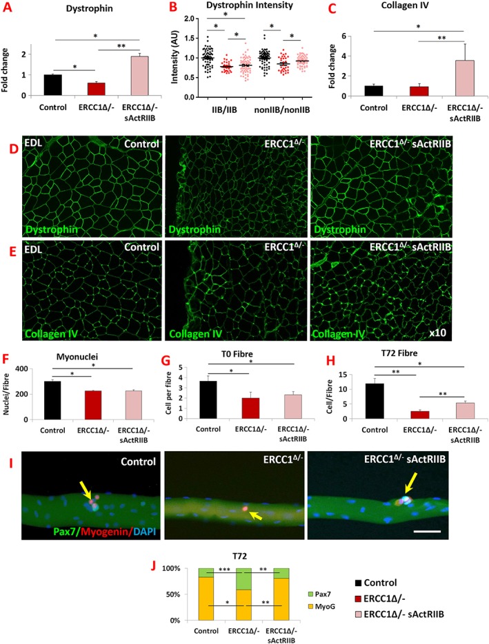
Figure 5.

Normalization of Ercc1 Δ/− extracellular components by sActRIIB and differentiation and self‐renewal of its satellite cells. (A) Dystrophin gene expression measured by quantitative PCR (qPCR). (B) Measure of dystrophin in fibre‐type‐specific manner using quantitative immunofluorescence. (C) Measure of collagen IV expression profiling by qPCR. (D) Immunofluorescence image for dystrophin expression in EDL muscle. (E) Immunofluorescence image for collagen IV expression in EDL muscle. n = 7 for all cohorts. (F) EDL myonuclei count. (G) Quantification of satellite cells on freshly isolated EDL fibres. (H) Quantification of cells on EDL fibres after 72 h culture. (I) Control, mock‐treated Ercc1 Δ/−, and sActRIIB‐treated Ercc1 Δ/− fibre examined at 72 h for expression of Myogenin (red) and Pax7 (green). Arrows indicated satellite cell progeny. (J) Quantification of EDL differentiated (Pax7−/Myogenin+) vs. stem cell (Pax7+/Myogenin−) after 72 h in culture. Fibres collected from three mice from each cohort and minimum of 25 fibres examined. Scale 50 μm. Non‐parametric Kruskal–Wallis test followed by the Dunn's multiple comparisons used for (A–C). Rest of data was analysed using one‐way analysis of variance followed by Bonferroni's multiple comparison tests. *P < 0.05, **P < 0.01, ***P < 0.001. EDL, extensor digitorum longus; sActRIIB, soluble activin receptor type IIB.
