
Figure 4.
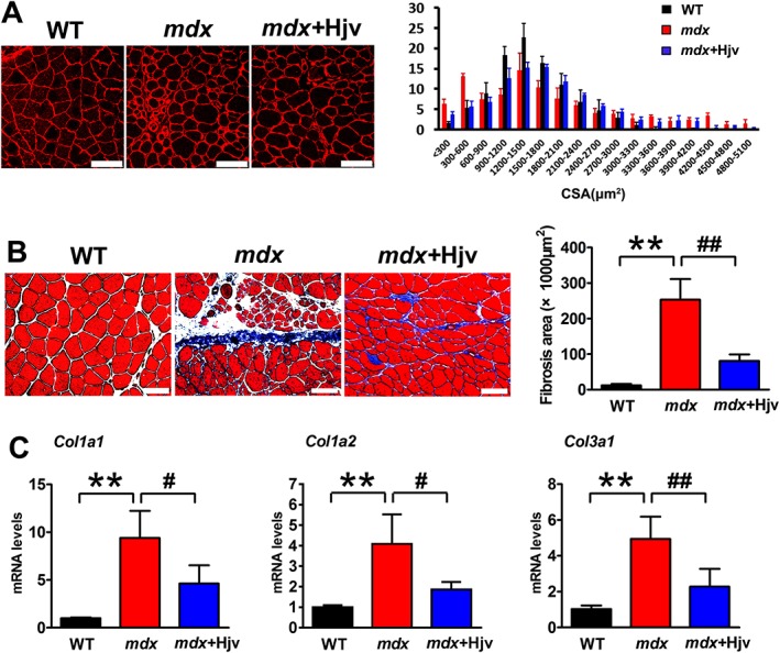
Hjv overexpression ameliorates dystrophic muscle wasting. (A) Immunostaining for laminin in muscle cross sections of WT and mdx mice transfected with control (mdx) or Hjv overexpression (mdx+Hjv) plasmids and the distribution plot of myofibre cross‐sectional area (n = 3). Scale bars: 100 μm. (B) Masson's trichrome staining and quantification of fibrosis area in muscle cross sections from the transfected muscles as shown in (A) (n = 4). Scale bars: 100 μm. (C) qRT‐PCR analysis of Col1a1, Col1a2, and Col3a1 mRNA levels expression in the transfected muscles shown in (A) (n = 4). All data are shown as mean ± SD. ** P < 0.01, # P < 0.05, ## P < 0.01 by one‐way analysis of variance. Hjv, hemojuvelin; WT, wild‐type.