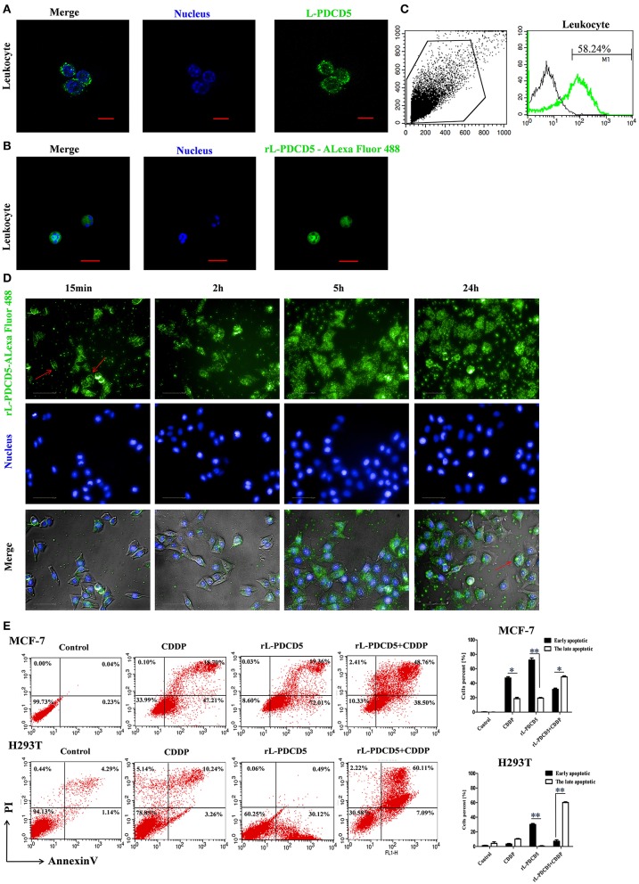
Figure 5.
The effect of rL-PDCD5 on morphological changes and apoptosis of leukocytes, MCF-7 cells, and H293T cells. (A) Immunofluorescence detection of L-PDCD5 in leukocytes. The primary antibody was the rabbit anti-rL-PDCD5 antibody (5.0 μg/mL), and the secondary antibody was an Alexa Fluor 488-conjugated goat anti-rabbit IgG antibody (5.0 μg/mL). The cells were observed and imaged with a Zeiss LSM 780 inverted microscope (Carl Zeiss, Inc.,) and analyzed using Zeiss ZEN LE software (60 × magnification). Scale bars: 5 μm. (B) Immunofluorescence detection of leukocytes treated with Alexa 488-labeled rL-PDCD5. The leukocytes were incubated in medium containing Alexa 488-labeled rL-PDCD5 (1.0 μg/μL) for 24 h and then incubated with Hoechst 33258 nucleic acid stain for 4 min. (C) FACS analysis of the efficiency of Alexa 488-labeled rL-PDCD5 entry into leukocytes. The leukocytes were incubated in medium containing Alexa 488-labeled rL-PDCD5 (1.0 μg/μL) for 24 h and then analyzed by FACS. (D) High-content screening analysis of MCF-7 cells treated with Alexa 488-labeled rL-PDCD5. MCF-7 cells were incubated with Alexa 488-labeled rL-PDCD5 (1.0 μg/μL) in a time-dependent manner and then incubated with Hoechst 33258 nucleic acid stain for 4 min. The cells were observed using high-content screening and photographed at the indicated time points (40 × magnification). (E) MCF-7 and H293T cells were exposed to the rL-PDCD5 (0.3 μg/μL) protein alone for 24 h or in combination with CDDP (20 μmol/L) for 6 h at 37°C. Apoptosis was analyzed by Annexin V/FITC and PI staining followed by flow cytometry. A histogram shows the statistics of the above mentioned results. The data are presented as the means ± SDs; n = 3, *P < 0.05, and **P < 0.01.

