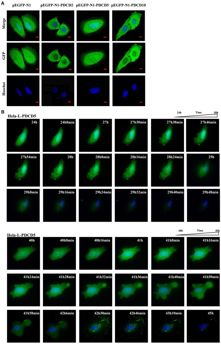
Figure 6.
Confocal microscopy and live cell imaging of the overexpression of L-PDCD5 in HeLa cells. (A) Confocal microscopy observation of HeLa cells transfected with L-PDCD2, L-PDCD5, and L-PDCD10 for 24 h and then treated either with or without CDDP (103 μmol/L) for 6 h. Hoechst 33342 was used to stain the nucleus. (B) Time-lapse imaging of L-PDCD5 overexpression in living cells (also shown in Supplemental Video 1). HeLa cells were transfected with L-PDCD5 for 24–45 h and stained with Hoechst 33342. The bubbling phenomenon was first observed in the cells, and the cells subsequently aggregated with each other until a small aggregate/particle-like structure was formed.

