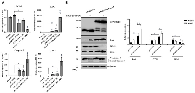
Figure 8.
The effect of the exogenous overexpression of L-PDCD5 on apoptosis-related proteins. (A) Q-PCR analysis of apoptotic molecule (BAX, BCL-2, TP53, and Caspase 3) expression in H293T cells transfected with L-PDCD5 for 24 h and treated either with or without CDDP (103 μmol/L). Total RNA was quantified by Q-PCR and normalized to GAPDH expression. All experiments were repeated at least three times with similar results (*P < 0.05, **P < 0.01, #P < 0.05, and ##P < 0.01). (B) Western blotting analysis of BAX, BCL-2, TP53, and Caspase 3 protein expression using specific antibodies. β-actin was used as a loading control (left side). A histogram showing the statistics of the above mentioned results (right side). All experiments were repeated at least three times with similar results (*P < 0.05, **P < 0.01, #P < 0.05, and ##P < 0.01).

