Figure 2.
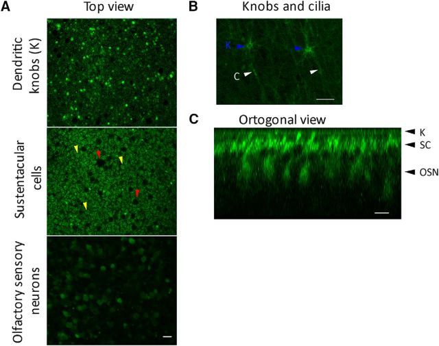
Uptake and accumulation of glucose analog by the cilia and dendritic knobs. A, Three sequential optical sections starting from apical surface (top) in a whole-mount rat olfactory mucosa incubated with the fluorescent glucose analog 2-NBDG in the bathing solution. The bright dots of the top and central images correspond to dendritic knobs; in the central image SC somata (yellow arrowheads) and Bowman's glands ducts (dark spots, red arrowheads) are appreciated; and OSN somata are distinguished in the bottom image. B, Higher-magnification view of the epithelial surface, showing that both, knobs (blue arrowheads) cilia (white arrowheads) incorporated the glucose analog. C, Orthogonal view of the epithelium reconstructed from a stack of images as in A, showing the different cell types; arrowheads on the right indicate the planes used in A, where OSN knobs, dendrites, and cell bodies, and SCs can be observed. Scale bars, 10 μm.

