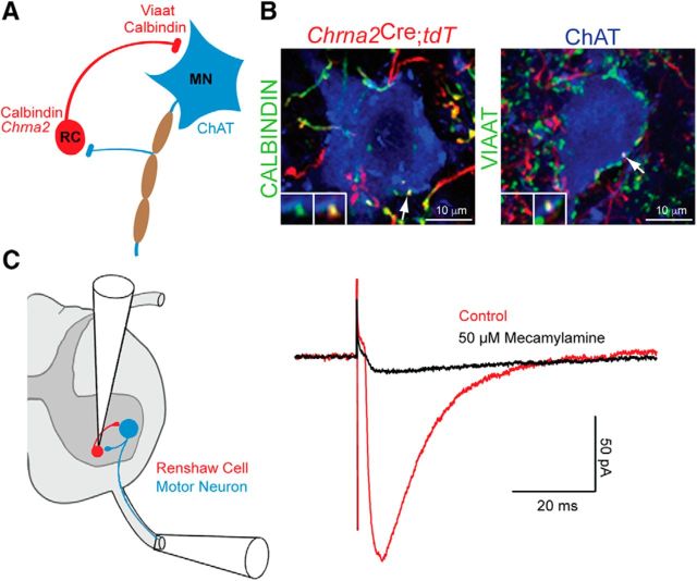
Figure 1.
Chrna2Cre labels Renshaw cell-derived synapses on motor neurons. A, Schematic illustration of the recurrent inhibitory (RC–MN) circuit. Calbindin and Chrna2+ Renshaw cells (red) form inhibitory VIAAT synapses on motor neurons and are reciprocally innervated by cholinergic motor neuron axons (blue). B, Immunohistochemistry of calbindin+ (left) and VIAAT+ (right) contacts derived from Chrna2Cre; R26.lsl.tdTomato+ cells on motor neurons (ChAT) in the lumbar spinal cord of adult mice. Inset shows the overlap among VIAAT, calbindin, and Chrna2Cre; R26.lsl.tdTomato. C, A schematic illustration of a hemisected spinal cord slice detailing the antidromic ventral root setup (left) with example traces showing the response of a Chrna2Cre; R26.lsl.tdTomato+ cell to ventral root stimulation in the absence (red) and presence (black) of mecamylamine (right).

