Figure 6.
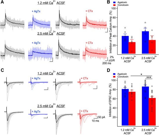
Ca2+ influx through P/Q- and N-type VGCCs differently control basal glutamate release. A, Representative examples of Ca2+ elevations recorded in MF boutons using Fluo-5F before and following application of AgTx or CTx, in both external Ca2+ concentrations. Individual traces correspond to individual points recorded simultaneously in MF boutons with the average overlaid. Traces are the average of 20 trials. B, Bar graph showing the inhibition of peak Ca2+ transients' amplitude by AgTx and CTx as a function of the external Ca2+ concentration. The relative contribution of P/Q- and N-type VGCCs to bouton-averaged Ca2+ elevations is not affected by the external Ca2+ concentration. Squares show data from individual boutons. C, Representative examples of MF-evoked EPSCs recorded in CA3 pyramidal cells in control conditions and in the presence of AgTx (blue) or CTx (red). The external calcium concentration was set to 1.2 or 2.5 mm. Traces shown are 20 consecutive trials and their average. D, Bar graph showing the percentage of EPSC inhibition by AgTx and CTx as a function of the external Ca2+ concentration. Squares show individual neurons. Data represent mean ± SEM. *p < 0.05, ***p < 0.001.

