Figure 7.

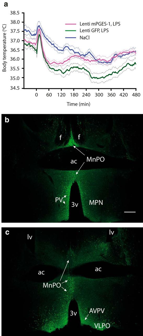
Mice injected into the preoptic hypothalamus with lentiviral vector encoding the terminal prostaglandin E2-synthesizing enzyme mPGES-1 display higher body temperature in response to LPS injection than mice injected with control vector. a, Temperature recordings of immune-challenged mPGES-1 knock-out mice injected with a viral vector encoding mPGES-1 (magenta trace) or given control injection of lentiviral vector encoding GFP (green trace). n = 12 for lenti-mPGES-1 LPS; n = 10 for lenti-GFP LPS; and n = 10 for NaCl (mixed group of lenti-mPGES-1 and lenti-GFP-injected mice). Error bars = SEM. b, c, Micrographs showing immunofluorescent staining for GFP in the preoptic hypothalamus after injection with viral vector. b and c are from different animals; the plane of the chosen sections corresponds approximately to bregma +0.14/+0.145 mm (b), and bregma +0.26/+0.245 mm (c) in the atlas of Paxinos and Franklin (2001) and the Allen Reference Atlas (Dong, 2008), respectively. 3v, Third ventricle; ac, anterior commissure; AVPV, anteroventral periventricular nucleus; f, fornix; lv, lateral ventricle; MnPO, median preoptic nucleus; MPN, medial preoptic nucleus; PV, periventricular nucleus; VLPO, ventrolateral preoptic nucleus. Scale bar, 500 μm.
